
A Positron Emission Tomography – Computed Tomography (PET-CT) scan combines two powerful imaging techniques – PET and CT scans. This imaging technique provides highly detailed 3-dimensional color images of what is happening inside a patient’s body at the molecular and cellular level.
In this nuclear medicine procedure, a small amount of radioactive glucose (typically fludeoxyglucose-18, similar to natural sugar) is injected into a vein. This radioactive tracer helps highlight the abnormal metabolic activity, thus assisting the healthcare providers in examining how tissues and organs are working. It is also useful in guiding the treatment of other medical conditions like Alzheimer’s disease. Given the rapid advancement in PET and CT technologies, there has been a considerable improvement in spatial resolution and sensitivity, thus enabling faster image acquisition and reducing radiation exposure for patients.
Why this test is done?
A PET CT scan is a useful tool in the diagnosis and evaluation of cancer. By evaluating organs and/or tissues for the presence of disease or other conditions, the scan provides more insight into the organ’s functionality. The scan may also be used to detect and monitor conditions such as:
- Parkinson’s disease. A progressive disorder of the nervous system
- Huntington’s disease. A hereditary disease of the nervous system
- Dementias: This condition causes deterioration of cognitive and mental function
- Cerebrovascular accident (stroke)
- To determine the function of the brain following trauma and evaluate hematoma (blood clot), bleeding, and/or perfusion
- Epilepsy. A brain disorder involving recurrent seizures
- To evaluate myocardial perfusion (blood flow to the heart muscle)
- To determine lung lesions or masses
- To detect tumour recurrence earlier than with other diagnostic modalities
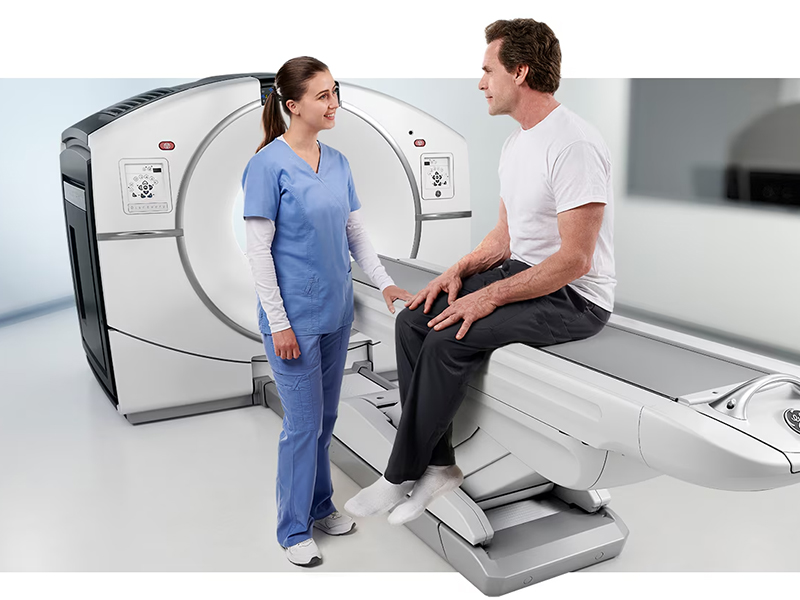
PET CT Scan procedure
Before the scan:
For this scan, the patient will need to stop eating and drinking (except water) at least six hours prior the scan. They can take regular medications with water, except diabetic medications. They should also avoid strenuous activity (such as aerobics or jogging) at least 24 hours before the scan. Diabetic patients should stop the oral hypoglycemic medications that contain Metformin 48 hours before the scan.
To know “pet ct scan price” in Delhi-NCR, do an online search to know the best pricing offered by diagnostic centres.
During the scan:
The patient will need to rest quietly, avoiding movement and talking. Depending on the type of nuclear medicine exam the patient is undergoing, a small amount of a radiotracer such as fluorodeoxyglucose (FDG), which contains both a sugar and a radioactive element, is injected. The radiotracer travels through the body and is absorbed by tumours or cancer cells. The patient then lies on the examination table, which moves slowly into the PET/CT scanner.
The imaging process will then begin. Following the PET scan, the CT scan will follow. While the PET scan takes 20 to 40 minutes, the CT scan takes less than two minutes. The total scanning time varies from 20 to 40 minutes. Depending on the organ or tissue being examined, additional tracers or medications may be administered. These imaging scans may help clinicians to gain a better insight into where the disease is occurring, how aggressive it is, and how well the the treatment is working. This may reduce the need for unnecessary surgery and help guide the ongoing therapy. Depending on the course of treatment selected by physicians, patients may require several scans during the course of their disease.
Once the scan is over, A trained radiologist or nuclear medicine physician will review the images. These images will then be interpreted and a detailed report will be prepared for the physician who ordered the tests.
Disclaimer: For personalised advice and further information, always consult your doctor or qualified healthcare professional.
